![]()
INTEGRATION OF A MULTIPLEXED ELECTROKINETIC PRECONCENTRATION CHIP INTO A MICROARRAY FOR RAPID AND ULTRASENSITIVE NUCLEIC ACID DETECTION
Jongmin Kim1, Ajymurat Orozaliev1, Sarah Sahloul1, Yong-Ak (Rafael) Song1, 2, 3, *
1Division of Engineering, New York University Abu Dhabi, PO Box 129188, Abu Dhabi, UAE
2Department of Chemical and Biomolecular Engineering, Tandon School of Engineering, New York University, NY 11201, USA
3Department of Biomedical Engineering, NYU Tandon School of Engineering, New York University, NY 11201, USA
*corresponding author: rafael.song@nyu.edu
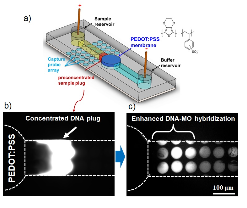
The Micro- and Nanoscale Bioengineering Group at NYU Abu Dhabi has developed a microfluidic electrokinetic concentrator chip in polydimethylsiloxane (PDMS) that can readily be interfaced with standard microarrays for high-throughput assays (see Figure 1a). It enhances the detection speed of bioassays by concentrating the target sample on microarrays. In this way, even a very low-concentrated analyte can be detected within a short amount of time.
After printing capture molecules (MO: morpholinos) on a glass substrate with iTWO, the PDMS concentrator was reversibly placed on top of the substrate. Once a voltage of 50V was applied across the channel, a concentration plug representing DNA was visible in the microchannel (see Figure 1b).
To quantify the concentration increase, we performed an experiment with three different concentrations of DNA ranging from 1 to 100 nM. In all cases, the concentration increased rapidly by more than two orders of magnitude in the first minute and then stabilized after about 5 min. The DNA concentration reached a stable value of ~0.3 μM (300-fold increase), ~4 μM (i.e. 400-fold increase), and 80 μM (800-fold increase) for 1 nM, 10 nM, and 100 nM, respectively.
The enhanced DNA-morpholino hybridization can clearly be seen between the concentrated and non-concentrated zone in the microchannel (see Figure 1c). The ease of integration of the concentrator device with standard microarray makes our preconcentration microfluidic chip a promising enhancing tool for microarray-based assays.
[1] Diogo Martins, Rastislav Levicky, Yong-Ak Song, Biosensors and Bioelectronics, Volume 72, 2015, pp 87–94
[2] Diogo Martins, Xi Wei, Rastislav Levicky, Yong-Ak Song, Analytical Chemistry, 2016, 88 (7), pp 3539–3547